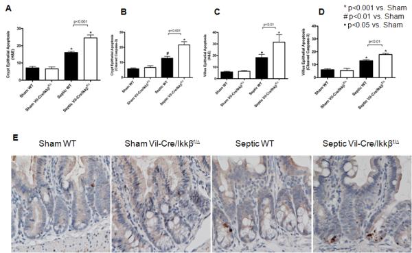
Figure 3.

Sepsis-induced intestinal epithelial apoptosis is exacerbated in mice lacking functional enterocyte NF-kB. Intestinal apoptosis was quantified in crypt (A and B) and villus (C and D) epithelium via H&E staining (A and C) and active caspase-3 staining (B and D). Septic WT mice exhibited increased epithelial apoptosis compared to shams by both methods, while septic Vil-Cre/Ikkßf/Δ mice exhibited exacerbated epithelial apoptosis compared to septic WT mice. n=9-12/group. Representative histomicrographs (E) are shown for sham and septic WT and Vil-Cre/Ikkßf/Δ mice stained for active caspase 3. Active caspase 3 staining in the crypt epithelium appears brown.
