
Figure 4.
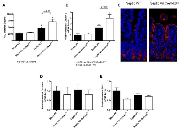
Sepsis-induced intestinal hyperpermeability is exacerbated in mice lacking functional enterocyte NF-kB. Intestinal permeability was evaluated in vivo by measuring the amount of FITC-conjugated dextran (FD-4) in the plasma (A). Septic WT mice exhibited increased permeability compared to shams, while septic Vil-Cre/Ikkßf/Δ mice had a further increase in intestinal permeability compared to septic WT mice. n=9-12/group. Gene expression of claudin-2 (B) was then evaluated by qRT-PCR. Claudin-2 levels were increased in septic WT mice compared to shams, while septic Vil-Cre/Ikkßf/Δ mice had a further increase in claudin-2 expression. n=8-14/group. Localization of claudin-2 was evaluated by fluorescent immunohistochemistry (C). Compared to sham WT and septic Vil-Cre/Ikkßf/Δ mice which had minimal staining (not shown), claudin-2 expression was increased in septic WT mice and localized predominantly in the crypts. In contrast, septic Vil-Cre/Ikkßf/Δ mice exhibited increased claudin-2 that was localized along the apical membrane of the villi as well as in the crypts. Representative images for each group are shown Magnification x20. Gene expression of occludin (D) and ZO-1 (E) was also evaluated by qRT-PCR. No differences were detected in either occludin or ZO-1, independent of whether an animal was subjected to CLP or had enterocyte NF-kB n=8-14/group.