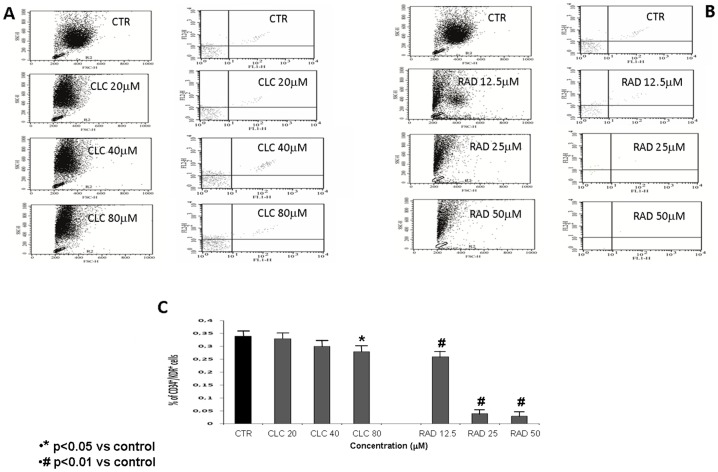
Figure 1. FACS analysis of samples of PBMNC derived from human healthy donors (see “Materials and Methods”) labelled with mouse monoclonal antibodies conjugated with fluorophores (CD34-FITC and KDR-PE).
Quantitative fluorescence analysis was performed with a FACS-CANTO instrument (BD Biosciences). Cells positive for both CD34-FITC and KDR-PE were considered as EPC. Isolated PBMCs incubated with CLC (20, 40 and 80 µM) (Fig.1 A) or RAD (12.5, 25 and 50 µM) (Fig.1 B) for 72 h in complete media at 37 C. Control cells were also cultured in complete media for 72 h at 37 C. Fig. 1 C shows the CD34+/KDR+ cells after treatment with CLC (20, 40 and 80 µM (p<0.05)) or RAD (12.5, 25 and 50 µM) (p<0.01) expressed as % of control. The figure is representative of three different experiments that always gave similar results.

