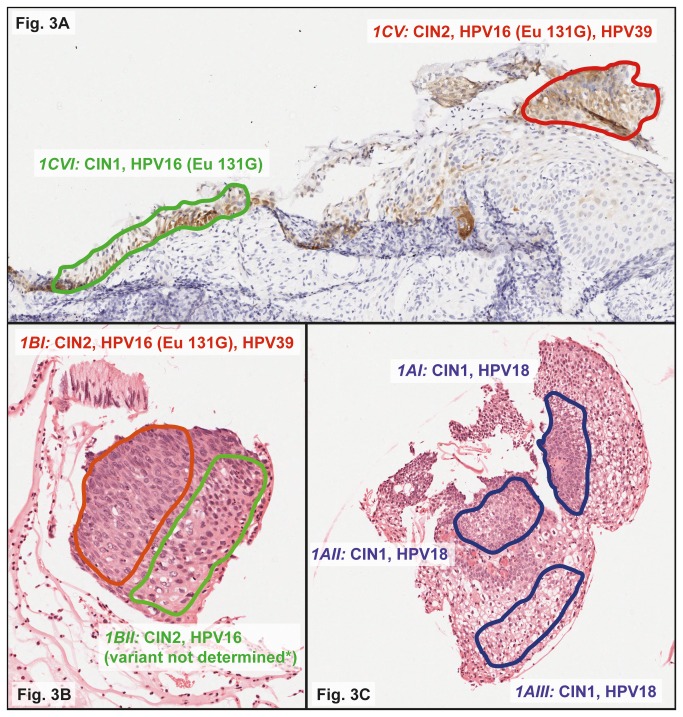
Figure 3. Histological images of localized HPV analysis by laser-capture micro-dissection (LCM) on a whole tissue section (WTS) positive for HPV16 (Eu 131G), 18 and 39.
Local excision by LCM was performed on colored regions of different grades of cervical intraepithelial neoplasia (CIN), i.e., 1CV and 1CVI from the p16-stained section (Figure 3A), and 1BI, 1BII (Figure 3B), 1AI, 1AII, and 1AIII (Figure 3C) from the hematoxylin and eosin (H&E)-stained section. Excised regions were separately analyzed by LiPA25 and subsequently by the HPV16 variant RHA if HPV16-positive. All Images have been captured by ScanScope XT digital scanner (Aperio Technologies Inc, Vista, Ca, USA). * Region 1BII was positive by LiPA25 for HPV16 but negative by the HPV16 variant RHA.

