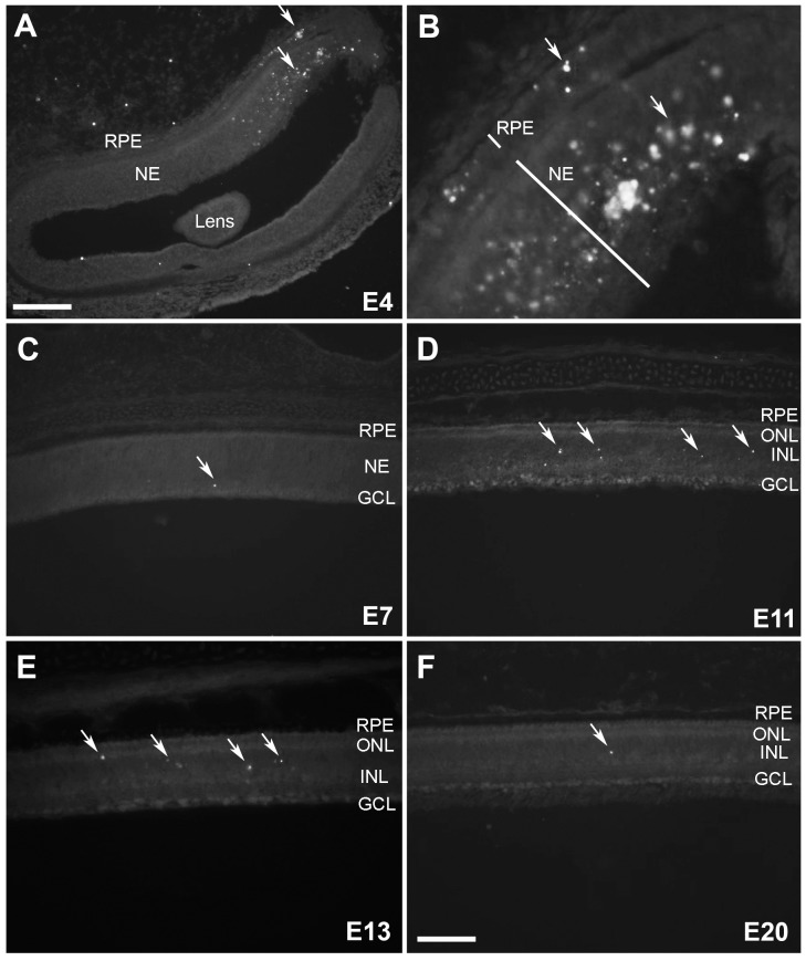
Figure 2. 5mC (+) cells in the developing chicken retina.
(A) The morphogenic furrow at E4 with a dense patch of 5mC staining (see arrows) (B) A magnified image of 5mC staining in the morphogenic furrow in a serial section taken at higher magnification. (C) 5mC (+) cells at E7, (D) E11, (E) E13 and (F) E20 are indicated by arrows when present. RPE = retinal pigment epithelium; NE = neuroepithelium; ONL = outer nuclear layer; INL = inner nuclear layer; GCL = ganglion cell layer. Scale bars = 75 µM in A and 200 µM in C–F.

