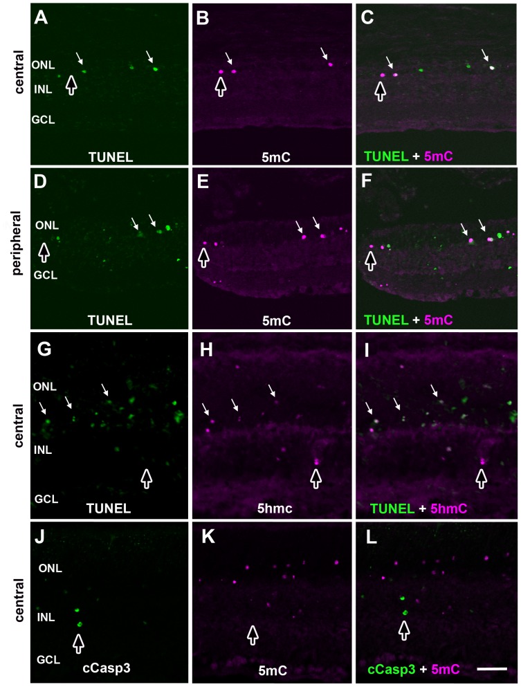
Figure 5. Cell death markers double labeled with 5mC (+) or 5hmC (+) photoreceptors in rd1 mouse retinas.
Retinas from P14 rd1 mice were double-labeled with TUNEL stain (A, D and G) and 5mC (B and E) or 5hmC (H) with central (A–C, G–I) and peripheral retina (D–F) shown accordingly. Discrete signals for cCaspase3 (J) and 5mC (K) labeling show virtually no overlap in signals (L). Overlap in signals are indicated by white arrows as shown in panels C, F and I. Cells labeled with one marker but not the other are indicated with dark vertical arrows. ONL = outer nuclear layer; INL = inner nuclear layer; GCL = ganglion cell layer. Scale bars = 50 µM.

