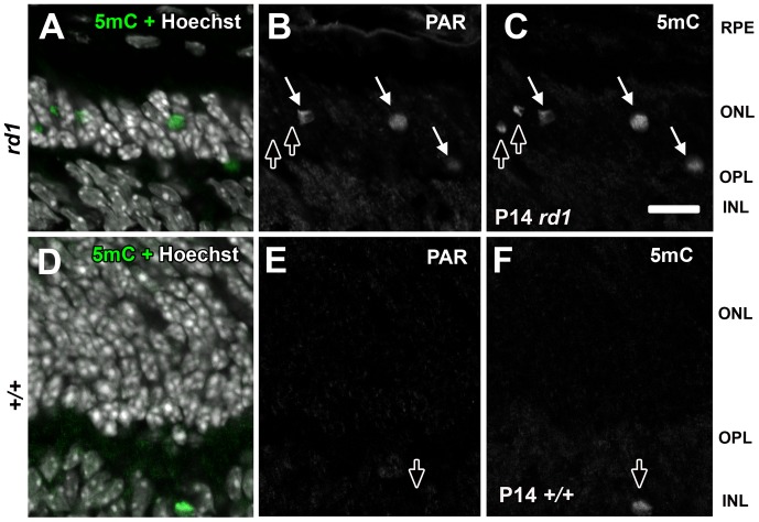
Figure 6. Detection of 5mC and PAR in the outer nuclear layer of degenerating rd1 retinas.
(A–C) P14 rd1 or (D–F) wild type mouse retinas were labeled with antibodies against 5mC and PAR. Panels A and D are co-labeled with Hoechst nuclear counterstain to indicate the respective retinal layer where 5mC exists. White arrows (B–C, E–F) indicate cells in which both antigens were detected. Dark arrows indicate cells that were labeled with 5mC but not PAR. RPE = retinal pigment epithelium, ONL = outer nuclear layer; OPL = outer plexiform layer, INL = inner nuclear layer; GCL = ganglion cell. Scale bar (L) = 50 µM.

