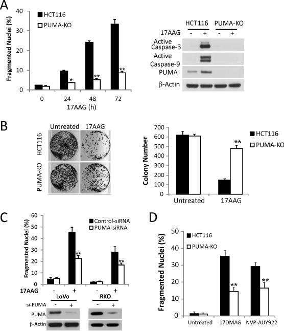
Figure 2. PUMA is required for Hsp90 inhibitor-induced apoptosis.
(A) WT and PUMA-KO HCT116 cells were treated with 1 μM 17AAG. Left, apoptosis at the indicated times was analyzed for by counting condensed and fragmented nuclei. **, P < 0.001, WT vs. PUMA-KO. Right, Active caspase-3 and caspase-9 at 72 h were analyzed by Western blotting. (B) Colony formation assay was done by seeding an equal number of WT and PUMA-KO HCT116 cells treated with 1 μM 17AAG for 48 h in 12-well plates, and the attached cells were stained with crystal violet after 14 days. Representative pictures of colonies (Left) and quantification of colony numbers (Right) are shown. **, P < 0.001, WT vs. PUMA-KO. (C) LoVo and RKO cells were transfected with either a scrambled siRNA or PUMA siRNA for 24 h and then treated with 1 μM 17AAG for 48 h. Top, apoptosis was analyzed by counting condensed and fragmented nuclei. **, P < 0.001, si-PUMA vs. Scrambled. Bottom, Western blotting confirmed PUMA depletion by siRNA. (D) WT and PUMA-KO HCT116 cells were treated with 0.25 μM 17DMAG and 0.5 μM NVP-AUY922 for 48 h. Apoptosis was analyzed by counting condensed and fragmented nuclei. **, P < 0.001, WT vs. PUMA-KO.

