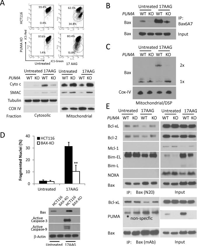
Figure 3. PUMA mediates 17AAG-induced apoptosis via the mitochondrial pathway and Bax activation in HCT116 cells.
The indicated cells were treated with 1 μM 17AAG or vehicle (untreated) for 48 h. (A) Upper, mitochondrial membrane potential was analyzed by flow cytometry following staining with JC-1. The circled lower populations indicate decreased red/green ratio and membrane depolarization. Lower, the distribution of Cytochrome c and SMAC in mitochondrial or cytosolic fraction was analyzed by Western blotting. α-Tubulin and Cytochrome oxidase subunit IV (COX IV) were used as the control for fractionation. (B) Bax conformational change was detected by immunopercipiation (IP) with anti-Bax 6A7 (activated) antibody followed by Western blotting. (C) Bax multimerization in isolated mitochondria was analyzed by Western blotting under non-denaturing conditions following DSP crosslink. (D) Apoptosis was analyzed by counting condensed and fragmented nuclei (left) and Western blotting of active caspase-3, caspase-9 (lower). β-action was used as the control for loading. (E) Interactions of endogenous Bax with indicated Bcl-2 family members were analyzed by Immunoprecipitation (IP) followed by Western blotting. The Rabbit (N20, upper panels) and mouse (mAb, lower panels) anti-Bax antibodies were used.

