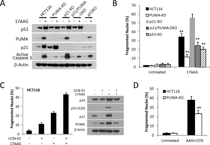
Figure 4. p53-dependent p21-induction suppresses 17AAG-induced apoptotic.
(A) HCT 116 cell lines with the indicated genotypes were treated with 1 μM 17AAG for 48 h. Western blotting of PUMA, p21, p53 and active caspase-3. (B) Apoptosis was analyzed by counting condensed and fragmented nuclei in the indicated cell lines at 48 h. **, P < 0.001, p21 KO vs. WT, p53-KO, PUMA-KO, or p21/PUMA-DKO. (C) HCT116 cells were treated with 1 μM UCN-01 or 1 μM 17AAG or their combination for 48 h. Left, apoptosis was analyzed by counting condensed and fragmented nuclei. Right, the expression levels of total p53, p-p53 (S20), p21 and PUMA were analyzed by Western blotting. (D) WT and PUMA-KO HCT116 cells were treated with 1 μM 17AAG and 1 μM UCN-01 for 48 h. Apoptosis was analyzed by counting condensed and fragmented nuclei. **, P < 0.001, WT vs. PUMA-KO.

