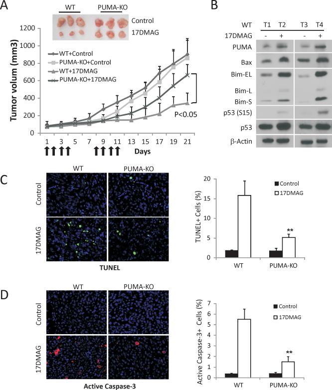
Figure 6. PUMA contributes to the anti-tumor activities of 17DMAG in a xenograft model.
(A) Nude mice after 1 wk of implantation of 4×10⁶ WT or PUMA-KO HCT116 cells were treated with 15 mg/kg of 17DMAG or the control buffer by i.p. injection on days 1-4 and 7-11. Tumor volume at indicated time points after treatment was calculated and plotted (n=6 in each group). Top inset, representative tumors at the end of the experiment. (B) HCT116 WT xenograft tumors from mice treated with 15 mg/kg of 17DMAG or the control buffer for five consecutive days. The indicated proteins were analyzed by Western blotting in representative tumors. (C) Paraffin-embedded WT and PUMA-KO tumor sections from mice treated as in (B) were analyzed by TUNEL staining. Left, representative TUNEL staining pictures; Right, TUNEL positive cells were counted and plotted. (D) Paraffin-embedded WT and PUMA-KO tumor sections from mice treated as in (B) were analyzed by active caspase-3 staining. Left, representative staining pictures. Right, active caspase-3 positive cells were counted and plotted.

