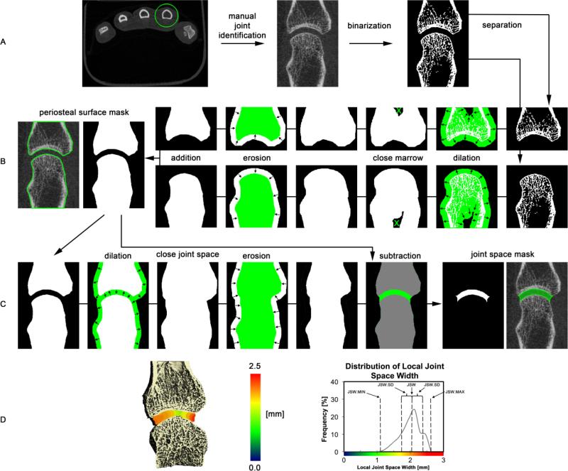
Figure 1.
Schematic overview of the image processing steps for the joint space segmentation. In the first step (A), all mineralized bone structures are extracted by thresholding and the individual bones are segmented using the relative supero-inferior position (MCP) or bone size and supero-inferior position (wrist). In the following step the periosteal surfaces are individually identified for each bone in the joint of interest (B). Next, the joint space volume is identified by closing the joint space using dilation/erosion of the juxta-ariticular surfaces followed by subtraction of the original volume contained within the periosteal surface (C). Finally, joint space morphometric parameters are derived from the 3D map of local joint space widths, shown in pseudo-color here for the second MCP of a subject with RA, and corresponding distribution of local joint space widths.

