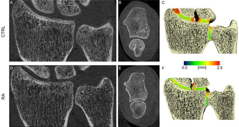
Figure 5.
Representative tomographic 2D coronal and axial images of the wrist (left and middle respectively), and 3D surface reconstructions of the wrist with the local JSW mapped into the joint space in pseudo-color (right). Shown is a healthy 25 year old individual (A-C) and a RA subject with various joint space abnormalities, including global joint space narrowing and joint space heterogeneity (D-F).

