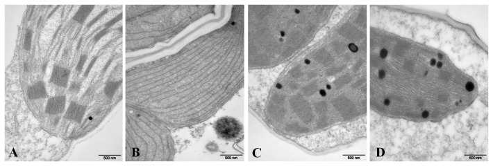
Figure 4. Electron microscope images of chloroplast structures.

A and C, Zheng58 wild-type plants at the seedling and tasseling stages, respectively; B and D, elm2 mutant plants at the seedling and tasseling stages, respectively. Scale bar=500 nm.

A and C, Zheng58 wild-type plants at the seedling and tasseling stages, respectively; B and D, elm2 mutant plants at the seedling and tasseling stages, respectively. Scale bar=500 nm.