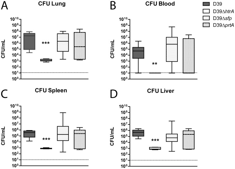
Figure 2. S. pneumoniae D39ΔhtrA, but not D39Δsfp or D39ΔprtA, displays diminished growth and dissemination in vivo.
Mice were infected with WT or mutant S. pneumoniae (5×10⁵ CFU) via the intranasal route and euthanized 48 hours later. Bacterial counts were determined in lung (A), blood (B), spleen (C) and liver (D). Data are expressed as box- and whisker plots depicting the smallest observation, lower quartile, median, upper quartile and largest observation. N = 8 mice per group at each time point. *** P < 0.005 versus WT S. pneumoniae.

