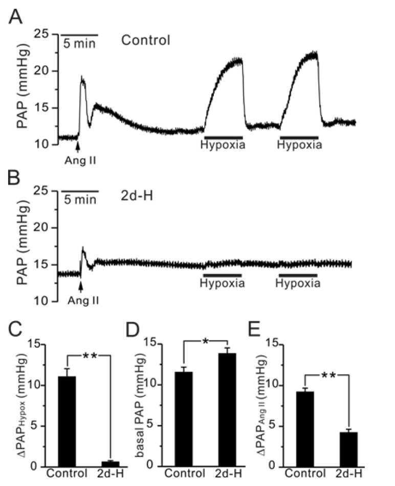
Fig. 1.
Comparison of HPV, basal PAP and Ang II-induced ΔPAP between control and 2d-H rats in V/P lungs. (A, B) Representative traces of PAP recording in control (A) and 2d-H rats (B). Hypoxic ventilation (3% PO2)-induced PAP increase (ΔPAPHypox) was abolished in 2d-H. Ang II was initially applied to the perfusate, inducing a transient increase in PAP. (C~E) Summaries of ΔPAPHypoxia (C), basal PAP (D), and AngII-induced ΔPAP (E) are shown as bar graphs comparing the results between control and 2d-H rats (*p<0.05, **p<0.01).

