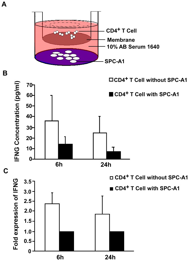
Figure 3. Suppressed IFNG expression of CD4+ T cell in the SPC-A1 transwell culture system.
(A) CD4+ T cells co-cultured with SPC-A1. CD4+ T cells (6×10⁵ cells/well) and SPC-A1 (2×10⁵ cells/well) were grown separated by a 0.4 µm pore insert in 24-well culture plates. SPC-A1 cells were grown in the outer wells and CD4+ T cells were grown in suspension in the inner wells. (B) Production of IFNG in CD4+ T cells cultured with or without SPC-A1. Supernatants were collected after anti-CD3, or CD28 stimulation for 6 or 24 h, and IFNG detected by ELISA. Results are from experiments performed on CD4+ T cells from 6 healthy volunteers (error bars, SD). (C) Quantitative RT-PCR analysis of IFNG transcripts from CD4+ T cells cultured with or without SPC-A1. RNA was isolated after anti-CD3, or CD28 stimulation for 6 or 24 h. Results are from experiments performed on CD4+ T cells from 6 healthy volunteers. IFNG levels increased 2.37 fold when stimulated for 6 h in CD4+ T cells grown without SPC-A1 compared with CD4+ T cells grown with SPC-A1.

