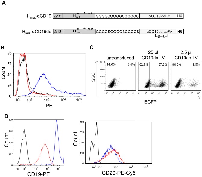
Figure 1. A stabilizing disulfide bond in αCD19ds-scFv is essential for generating CD19 targeting vectors.
(A) Schematic drawing of the cytoplasmic tail-truncated hemagglutinin envelope proteins displaying two αCD19 single-chain antibody (scFv) variants. In the mutated hemagglutinin protein (Hmut) that is derived from the NSe variant of the measles virus (MV) vaccine strain Edmonston B, mutations in the MV receptor recognition regions Y481A, R533A, S548L and F549S (ectodomain) [12] are indicated by asterisks. A glycine-serine linker ((G4S)3) is used as linker region between Hmut and scFv. The αCD19-scFv and αCD19ds-scFv differ in the presence of a disulfide bond in αCD19ds-scFv. A histidine tag (H6) is present at the scFv C-terminus. Both hemagglutinin proteins are truncated by 18 amino acids in their cytoplasmic tail (Δ18) to allow incorporation into the lentiviral envelope. (B) Cell surface transport of the two different H variants was investigated upon transfection of plasmids pCG-Hmut-αCD19 (red line) and pCG-Hmut-αCD19ds (blue line) into HEK-293T cells. The empty expression plasmid pCG-1 (black line) was used as control. Forty-eight hours after transfection, the cells were stained with a PE-conjugated anti-His antibody to detect cell surface expression of the H constructs by FACS analysis. The respective histograms are shown. (C) To determine the titer of vector particles pseudotyped with Hmut-αCD19ds (CD19ds-LV), CD19+ Raji cells were transduced with serial dilutions of the concentrated vector particles. After 72 h, the percentage of EGFP+ cells was determined by FACS analysis. (D) To determine cell surface down-regulation of CD19 after CD19ds-LV transduction, CD19+/CD20+ Raji cells were transduced with CD19ds-LV vectors at an MOI of 2 or remained untransduced. Forty-eight hours later, untransduced cells were stained with PE-conjugated anti-CD19 (left; blue line) and PE-Cy5-conjugated anti-CD20 (right; blue line) antibody, or with the respective isotype controls (black lines). CD19ds-LV transduced cells were stained with PE-conjugated anti-CD19 (left; red line) and PE-Cy5-conjugated anti-CD20 (right; red line) antibody. Then, CD19 and CD20 cell surface expression was analyzed by FACS.

