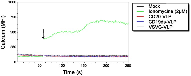
Figure 5. CD20-VLP and CD19ds-VLP do not induce calcium influx into unstimulated primary human B lymphocytes.

Freshly isolated primary human B lymphocytes were labeled with Calcium Sensor Dye eFluor® 514 and labeled cells were assessed by flow cytometry to establish a baseline level of fluorescence. Then cells were removed, incubated with the indicated stimulants, and replaced immediately for further flow cytometric analysis. The transient increase in intracellular calcium concentration was recorded by monitoring the change in MFI of the cells. To determine the background auto-fluorescence of the cells, one sample was recorded without the addition of any stimulant after baseline monitoring (mock). The gap in the histogram reflects the time-period when the tube containing the cells was removed from the instrument to add the stimulants. The arrow indicates the time-point of addition of the stimulant. VLP: virus like particles; MFI: mean fluorescence intensity.
