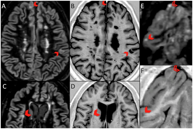
Figure 1. Corresponding DIR and PSIR images showing change of classification of CGM lesions.
DIR LC lesions (blocked chevron) in panel A, appear as JC-WM lesions on PSIR in panel B; DIR LC lesion in panel C is seen to be a pure IC lesion on PSIR in panel D; DIR LC lesion (blocked chevron) and IC lesion (open chevron) in panel E appear as JC WM and LC on PSIR respectively, in panel F.

