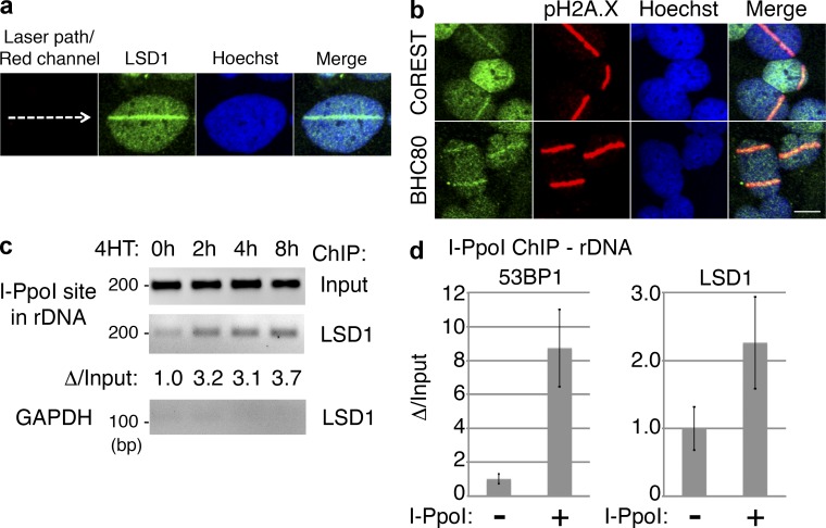
Figure 1.
LSD1 is recruited to sites of DNA damage. (a) UV laser microirradiation was performed on U2OS cells, incubated at 37°C for 10 min, stained for endogenous LSD1, and analyzed by confocal microscopy. (b) U2OS cells were microirradiated as in panel a, and then stained for CoREST or BHC80 and pH2A.X as indicated. Bar, 10 µm. (c) PCR was performed using rDNA or GAPDH primers with LSD1 ChIP material after DSBs were induced with retroviral I-PpoI in U2OS cells. The indicated time points represent the length of 4-OH-tamoxifen (4HT) treatment to induce DSBs. Shown below is the change of LSD1 rDNA ChIP quantified and normalized to input. (d) U2OS cells transduced with control or I-PpoI–expressing virus and were treated with 4HT for 4 h. ChIP was then performed using antibodies against 53BP1 or LSD1, and real-time qPCR was done on the ChIP material using rDNA primers.

