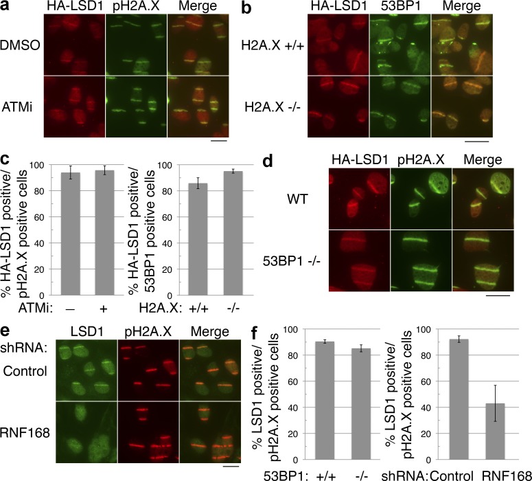
Figure 3.
LSD1 recruitment to sites of DNA damage is dependent on RNF168. (a) Flag-HA-LSD1 was stably expressed in U2OS cells, and then treated with the ATM inhibitor KU55933 (15 µM) or DMSO 1 h before laser microirradiation. The cells were then stained for HA and pH2A.X. (b) Wild-type (H2A.X+/+) or H2A.X-deficient (H2A.X−/−) MEFs stably expressing HA-tagged LSD1 were laser microirradiated and stained as in panel a. (c) Quantitation of a and b, with error bars representing the SD of duplicate experiments. (d) Wild-type or 53BP1-deficient (53BP1−/−) MEFs stably expressing Flag-HA–tagged LSD1 were laser microirradiated and stained as in panel a. (e) U2OS cells were treated with the indicated shRNAs, microirradiated, and stained for endogenous LSD1 and pH2A.X. (f) Quantitation of d and e, with error bars representing the SD of duplicate experiments. Bars, 20 µm.

