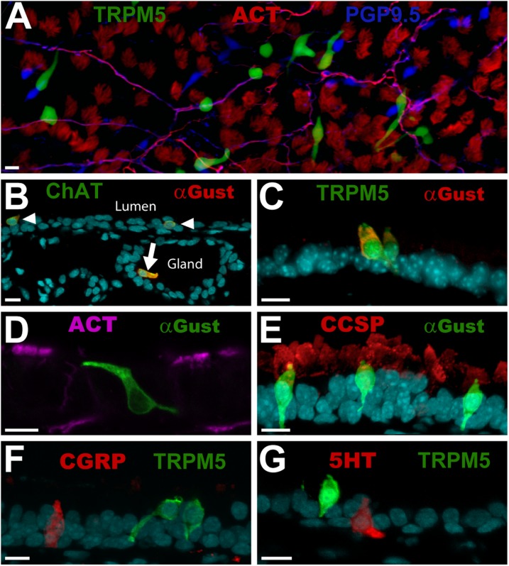
Figure 1.
Brush cells (BCs) comprise a distinct cell type in the tracheal epithelium. (A) Triple-labeled, whole-mount tracheal epithelium shows transient receptor potential melastatin 5 (TRPM5)–green fluorescent protein (GFP; green), protein gene product 9.5 (PGP9.5; blue), and acetylated tubulin (ACT; red). BCs appear green. Nerve fibers (magenta) are immunoreactive for both PGP9.5 and acetylated tubulin. Neuroendocrine cells are immunoreactive for PGP9.5 (blue), whereas cilia appear red, and are immunoreactive for ACT. (B) BCs coexpress Gα-gustducin (Gust; red) and choline acetyltransferase (ChAT; green), and occur within the tracheal epithelium (arrowheads) and in submucosal glands (arrow). (C) Most BCs are immunoreactive for both Gα-gustducin (red) and TRPM5 (green), and have multiple processes. (D) Gα-gustducin (green) immunoreactive BCs are not immunoreactive for ACT (magenta), a marker for ciliated cells. (E) Gα-gustducin (green) immunoreactive BCs are not immunoreactive for club (Clara)–cell secretory protein (CCSP) (red), a marker for club (Clara)–like cells. (F and G) TRPM5 (green)–expressing BCs appear morphologically similar to neuroendocrine cells, but are not immunoreactive for the known neuroendocrine cell markers (E) calcitonin gene related peptide (CGRP; red) and (F) 5-hydroxytryptamine (5HT; red). (B–G) Counterstains with DRAQ5 are shown in cyan. Photographs are of tissues from mice that ranged in age from 90–180 days. Scale bars = 10 μm.

