Figure 3.
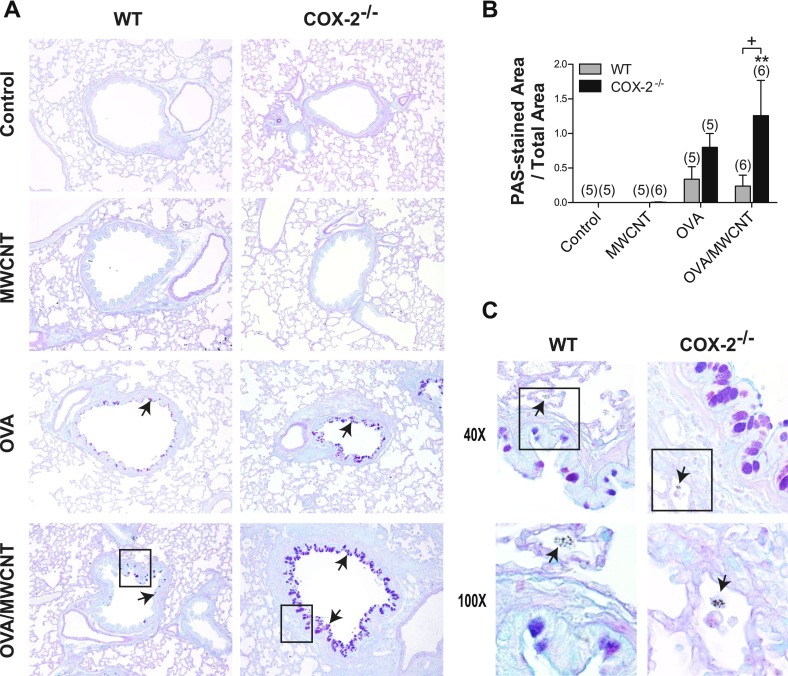
Mucus-cell hyperplasia after allergen and multiwalled carbon nanotube (MWCNT) exposure. (A) Photomicrographs of alcian blue–periodic acid–Schiff (AB-PAS)–stained sections of lung tissue taken 1 day after MWCNT exposure, low magnification (×10). Inset boxes are shown in higher magnification in C. Arrows indicate AB-PAS-positive mucus-producing cells in airways. (B) Semiquantitative scoring of AB-PAS–stained lung sections from mice, 1 day after exposure, shows that airway goblet cells (arrows) are significantly increased in the lungs of COX-2−/− mice after combined allergen challenge and MWCNT exposure, compared with WT mice. **P < 0.01, compared with COX-2−/− control mice. +P < 0.05, compared with wild-type (WT) ovalbumin (OVA)/MWCNT mice. Numbers in parentheses indicate number of animals in dosage group. (C) Higher magnification (×40) of inset from ×10 micrographs of OVA-sensitized, WT, and COX-2−/− mice exposed to MWCNTs, as well as higher magnification (×100) of inset from ×40 micrographs of OVA-sensitized, WT, and COX-2−/− mice exposed to MWCNTs. Arrows indicating alveolar macrophages containing MWCNTs are depicted in the photomicrographs at ×100.

