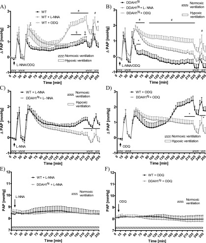
Figure 3.
Effects of Nω-nitro-l-arginine (L-NNA) and 1H-[1,2,4]oxadiazolo [4,3-a]quinoxalin-1-one (ODQ) on pulmonary arterial pressure (PAP) during normoxic and hypoxic ventilation of isolated lungs. (A) Increase of PAP (ΔPAP) during normoxic and hypoxic ventilation was referenced to PAP values at time point 0 (normoxic ventilation) in the presence of L-NNA and ODQ in isolated lungs from wild-type (WT) mice. *Significant difference (P < 0.05) between untreated WT control lungs and L-NNA–treated lungs. #Significant difference (P < 0.05) between untreated WT control lungs and ODQ-treated lungs. §Significant difference (P < 0.05) between ODQ-treated and L-NNA–treated lungs. No significant difference in ΔPAP was evident during the second acute hypoxic ventilation, performed at Minutes 235–245, when related to PAP values at the onset of hypoxic ventilation (235 minutes). Each data point is derived from n = 4–10 lungs per group. (B) Increase of PAP (ΔPAP) during normoxic and hypoxic ventilation was referenced to PAP values at time point 0 (normoxic ventilation) in the presence of L-NNA and ODQ in isolated lungs from DDAH1-overexpressing (DDAHtg) mice. *Significant difference (P < 0.05) between untreated WT control lungs and L-NNA treated lungs. #Significant difference (P < 0.05) between untreated WT control lungs and ODQ-treated lungs. No significant difference in ΔPAP was evident during the second acute hypoxic ventilation, performed at Minutes 235–245, when related to PAP values at the onset of hypoxic ventilation (235 minutes). Each data point is derived from n = 5–8 lungs per group. (C) Increase of PAP (ΔPAP) during normoxic and hypoxic ventilation was referenced to PAP values at time point 0 (normoxic ventilation) in the presence of L-NNA in isolated lungs from WT and DDAHtg mice. *Significant difference (P < 0.05) between lungs of WT and DDAHtg mice. No significant difference of ΔPAP was evident during the second acute hypoxic ventilation performed at Minutes 235–245, when related to PAP values at the onset of hypoxic ventilation (235 minutes). Each data point is derived from n = 5–7 lungs per group. Data are derived from L-NNA–treated groups in A and B. (D) Increase of PAP (ΔPAP) during normoxic and hypoxic ventilation was referenced to PAP values at time point 0 (normoxic ventilation) in the presence of ODQ in isolated lungs from WT and DDAHtg mice. *Significant difference (P < 0.05) between lungs of WT and DDAH1tg mice. No significant difference in ΔPAP was evident during the second acute hypoxic ventilation, performed at Minutes 235–245, when related to PAP values at the onset of hypoxic ventilation (235 minutes). Each data point is derived from n = 4–5 lungs per group. Data are derived from the ODQ-treated groups in A and B. (E) PAP during 255 minutes of normoxic ventilation in the presence of L-NNA. Each data point is derived from n = 3 lungs per group. (F) PAP during 255 minutes of normoxic ventilation in the presence of ODQ. Each data point is derived from n = 3 lungs per group.

