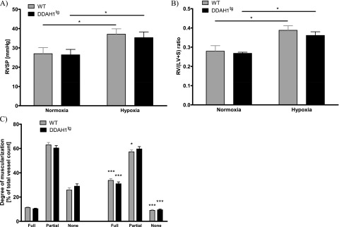
Figure 4.

Effects of DDAH1 overexpression on the development of hypoxic-induced pulmonary hypertension. (A) Right ventricular systolic pressure (RVSP) in normoxia and after 4-week exposure to hypoxia in wild-type (WT) and DDAH1-overexpressing (DDAH1tg) mice. *Significant difference (P < 0.05) between normoxia and hypoxia (n = 10 per group). (B) Ratio of right ventricular weight (RV) and left ventricular plus septal weight (LV + S) during normoxia and after 4 weeks of exposure to hypoxia in WT and DDAH1tg mice. *Significant difference (P < 0.05) between normoxia and hypoxia (n = 10 per group). (C) Degree of vascular muscularization is given as portions of fully, partly, and not muscularized vessels in percentages [%] of total vessels during normoxia and after 4 weeks of exposure to hypoxia in WT and DDAH1tg mice. *P < 0.05 and ***P < 0.001, significant difference compared with the respective normoxic group and hypoxic group (n = 10 per group).
