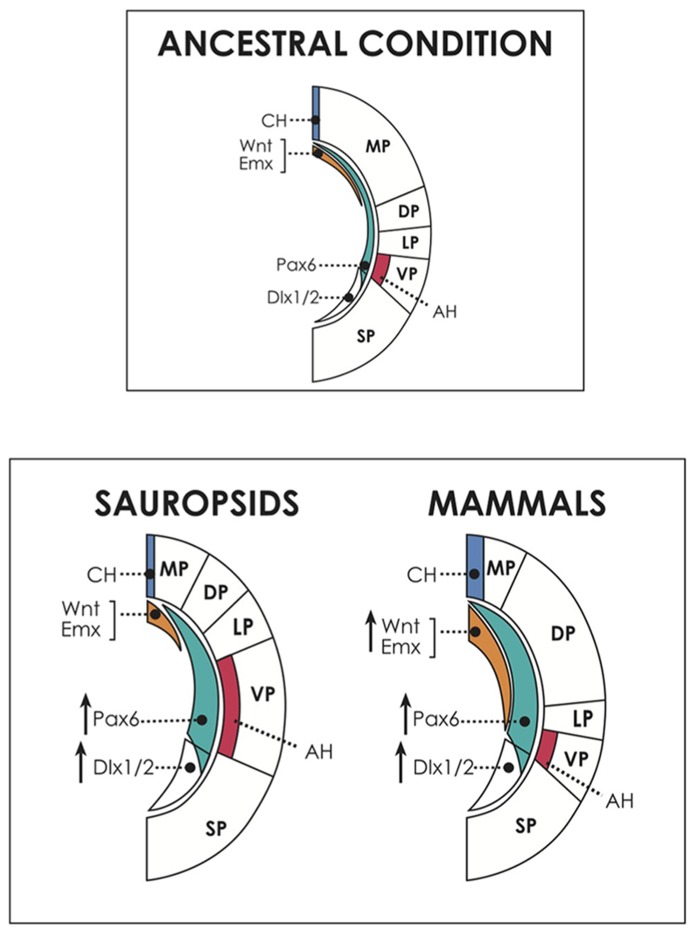
FIGURE 3.
Above, dorsal and ventral patterning centers in the cerebral hemispheres, and presumed ancestral condition. The dorsally located cortical hem (CH) expresses dorsalizing factors like Wnts and Emxs and patterns the embryonic medial pallium (MP, hippocampal formation and homologous structures) and the dorsal pallium (DP, neocortex in mammals; dorsal cortex/hyperpallium in sauropsids). On the other hand, the antihem, induced by Pax6 activity, specifies the ventral pallium (pallial amygdala in mammals, DVR/nidopallium in sauropsids). Pax 6 is expressed in a anteroventral-to-caudodorsal gradient that counteracts with the dorsalizing factors, and contributes also to neocortical and hippocampal patterning in mammals. In the common ancestor, perhaps similar of present-day amphibians, there was possibly a relatively large dorsal pallium, at the expense of the development of other pallial regions (Northcutt, 2013). Below, hypothetical scenario of developmental evolution in the pallium of amniotes. Pax6 is proposed here as a candidate to drive the amplification of progenitor proliferation in the brains of different amniotes, but there may be other or additional factors contributing to this process. Pax6 expression is proposed to have been upregulated in both sauropsids and mammals. In reptiles, this event produced a modest amplification of the antihem (AH) and the ventral pallium (VP), giving rise to the dorsal ventricular ridge. In birds, Pax6 amplification reached higher levels, expanding the nidopallium and mesopallium, and also reaching the DP, contributing to generate the hyperpallium. Conversely, in mammals, in addition to Pax6 enhancement there was a concomitant upregulation of dorsal signals (illustrated by an increase in Wnt and Emx activities), which antagonized Pax6 signaling, restricting the expansion of the antihem. Furthermore, in mammals, upregulation of Pax6 and dorsal signals show a significant overlap, allowing Pax6 to influence the expansion of the DP, giving rise to the neocortex. Not shown for simplicity is the anterior forebrain, patterned by the action of FGFs, which may have also contributed to brain expansion particularly in mammals. Note that the subpallium also increased in size in all amniotes. SP, subpallium, marked by the expression of markers like Dlx1/2.

