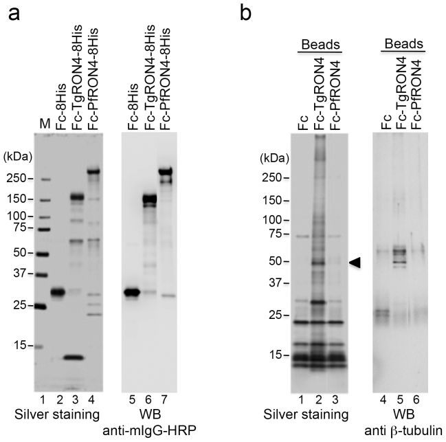
Figure 1. Identification of TgRON4-binding host proteins.
(a) Recombinant proteins expressed using the baculovirus expression system and purified on Ni-NTA agarose. Each protein (50 ng) was separated by 5%–20% gradient SDS-PAGE and subjected to silver staining (lanes 2–4). The molecular masses (kDa) are indicated on the left. Immunoblotting with an HRP-conjugated anti-mouse Fc antibody of purified recombinant proteins (lanes 5–7). (b) Each Fc-fusion protein was crosslinked to protein G magnetic beads and incubated with membrane proteins from 293 T cells. The eluate was separated by SDS-PAGE followed by silver staining (lanes 1–3). The arrowhead indicates a 50-kDa band that is specific for incubation with TgRON4-linked beads. Immunoblotting of the eluates with an anti-β-tubulin antibody (lanes 4–6).

