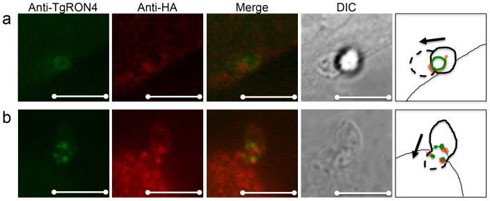
Figure 6. Localization of the TUBB2C C-terminal region and TgRON4 in the moving junction during Toxoplasma invasion.
CHO cells expressing HA-TUBB2C F3 were infected with T. gondii RH tachyzoites using a potassium buffer shift. At 2-min invasion, infected CHO monolayers were fixed and permeabilized. HA-TUBB2C F3 was stained with a rat anti-HA antibody and TgRON4 was stained with an anti-TgRON4 mouse polyclonal antibody. Alexa Fluor 350 goat anti-mouse IgG and Alexa Fluor 546 goat anti-rat IgG served as secondary antibodies. The HA-TUBB2C F3 accumulated around (a) or near (b) the moving junction. The cartoon illustrates the direction of the parasite invasion. Scale bar, 5 μm.

