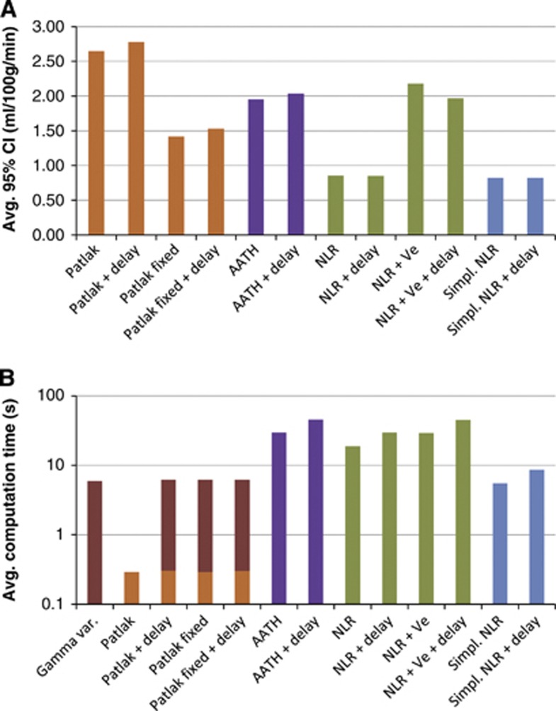
Figure 4.
(A) The average 95% confidence interval (CI) of Ktrans for of each method. More reliable estimates have narrower CIs. (B) The average computation per slice (512 × 512 pixels) for each of the methods on a low-end desktop PC. Note that the y axis is logarithmic. The Patlak methods that are extended with delay and/or fixed cerebral blood volume (CBV) require input from the gamma variate fit-based method. AATH, adiabatic approximation to the tissue homogeneity.

