Figure 6.

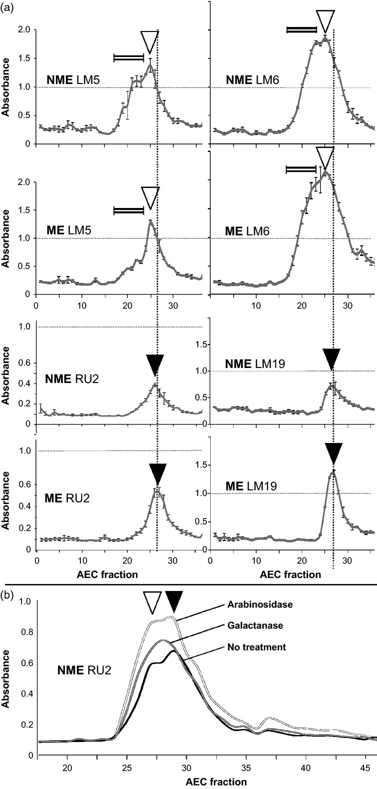
Epitope detection anion-exchange chromatography of extracts of the NME and ME halves of tobacco seed endosperms. Isolated cell-wall materials were fractionated using an anion-exchange chromatography column, and epitopes were detected in the same fractions by ELISA using LM5 galactan, LM6 arabinan, RU2 RG–I backbone and LM19 homogalacturonan (HG) monoclonal antibodies. Fractions indicated by the double-line symbol indicate the presence of early-eluting material using the LM5 and LM6 epitopes ahead of the peak of RG backbone and HG elution. Galactan-containing early-eluting peaks were more abundant in the NME than in the ME, and HG was more abundant in the ME than in the NME. Open triangles indicate coincident peaks of LM5 and LM6 epitope elution. Closed triangles indicate coincident peaks of RU2 and LM19 epitope elution. Vertical dotted lines indicate peaks of HG elution. Error bars indicate the SD of three absorbance values.
