Fig. 1.

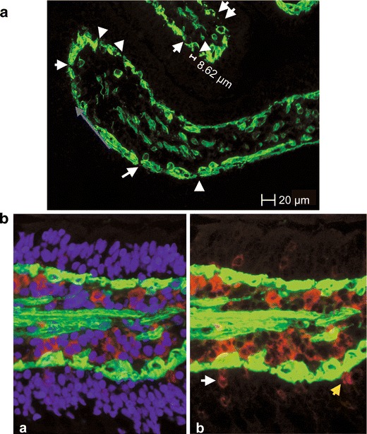
Evaluation of the in vivo experiment. a Laminin staining of the BM (green). Continuous staining was not detected, and the unstained areas of the basement membrane represent the pores of the BM. The length of the unlabelled area was taken as the diameter of the pores, given that the pores are circular. The blue arrow shows how the length of the BM was measured. b CD16+ cells (red) in close contact to the BM (laminin-stained, green); a triple-stained picture, nuclei (DAPI, blue), b CD16+ cells (white arrow) and a CD16+ dendrite (yellow arrow) in the epithelium
