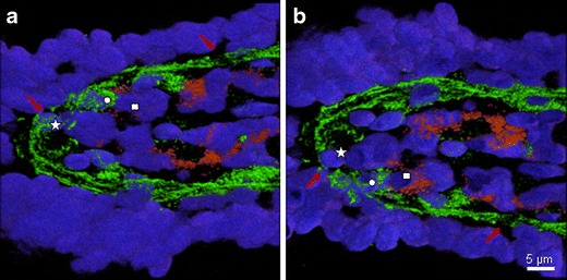
Fig. 5.

Three-dimensional reconstruction of the pores. Confocal microscopical pictures of a cryosection (16 μm) showing the laminin-labelled BM (green), CD16+ cells (red), and cell nuclei (DAPI, blue) studied. The optical sections were reconstructed and rendered to obtain the three-dimensional structure of the villus tip. Two views of the villus are given (a, b), the picture in (b) is rotated by 180° along the longitudinal axis. Symbols (square, star and circle) mark the position of the same three individual cells in both views. Red arrows mark characteristic pores. Two nuclei (star and circle) were found within a pore structure showing cells on the passage through the basal membrane. CD16+ cells were found in close contact to the basement membrane (see square for example)
