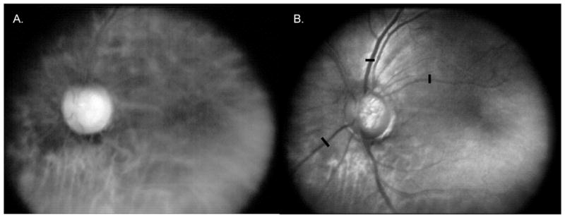
Figure 6.

Red illumination images of the same dilated 54 year old Caucasian Hispanic female patient as in Figure 5. A. DLP-Cam averaged red illumination dark field image, showing predominantly multiply scattered light from the deeper retina; B. Scaled difference between the averaged red illumination bright field image from Figure 5B and the dark field image shown in Figure 6A. The difference image is more confocal than the bright field image in Figure 5B, with greater observed vessel and optic nerve head contrast. This is confirmed by calculating the Michelson contrast across the retinal vessels at the same three locations indicated by the black bars.
