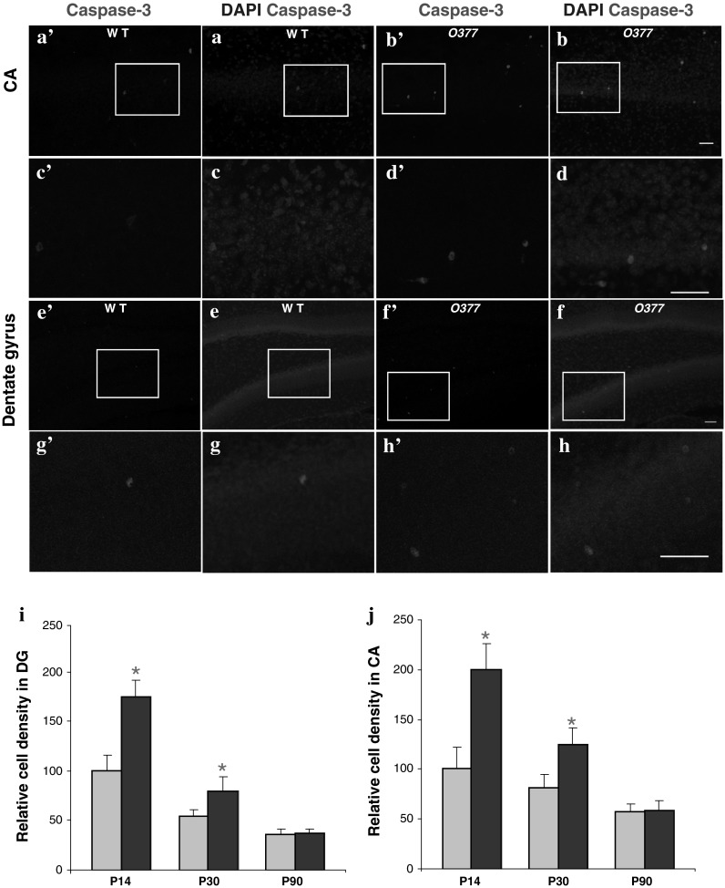
Fig. 2.

Apoptotic cell death is increased in young O377 mutants. a–h Confocal images of anti-caspase-3 staining (red) in 2-week-old wild-type (WT, left panel) and O377 mice (right panel) in the ventral CA (upper panel, a–d) and in the DG (lower panel, e–h). DAPI staining is given in blue. The boxed area is given below in a higher magnification. In i, j, the values are normalized to wild-type mice at the age of 3 months and given in % (T = 100 %). i Density of caspase-3-positive cells in WT and O377 mutants in the ventral DG (WT = 100 %). j Density of caspase-3-positive cells in wild-type and O377 mutants in the ventral CA (WT = 100 %). *p ≤ 0.05, Student’s t test, n ≥ 4. Scale bar 40 μm; error bar SEM
