
Figure 3.
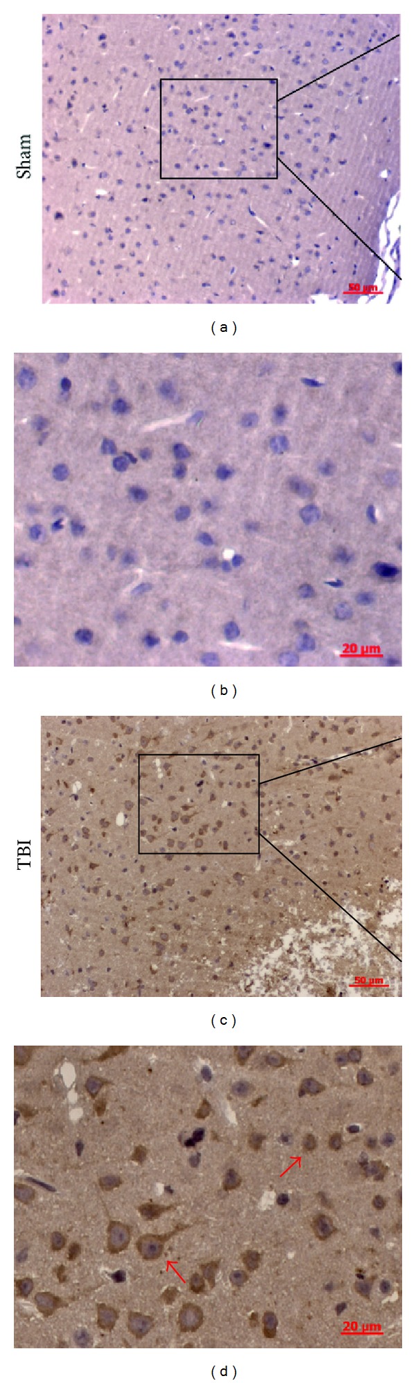
Representative photomicrographs showing cathepsin S (CatS) immunohistochemistry of tissue from the sham and traumatic brain injury (TBI) groups. In the sham group, almost none of the cells presented a positive morphology, whereas, in the TBI group, many cells were positive for CatS. CatS was present mainly in the cytoplasm. The CatS-positive cells in the TBI group (indicated by arrows) showed two populations of cells with different morphologies.