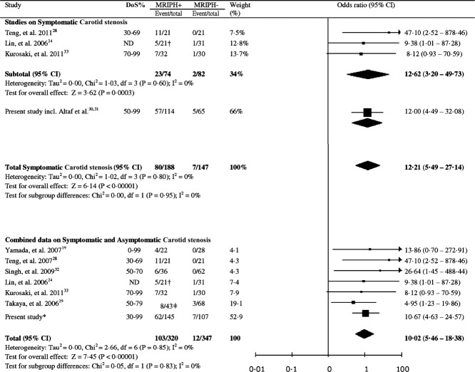
FIGURE 3.
Meta-analysis of available studies on symptomatic carotid arteries (n = 335), and symptomatic combined with asymptomatic carotid arteries (n = 667) to evaluate the association between magnetic resonance imaging (MRI) signal hyperintensity and future risk of ipsilateral cerebral ischemic events. *Combined data including symptomatic carotid artery stenosis and contralateral asymptomatic arteries. †Only included the subgroup of patients with MRI-defined intraplaque hemorrhage (PH) who were followed up for subsequent ischemic events. ‡PH+ within lipid-rich necrotic core plaque (LRNC) compared with PH− with LRNC; DoS = Degree of stenosis. CI = confidence interval; ND = not disclosed in the paper.

