Fig. 3.
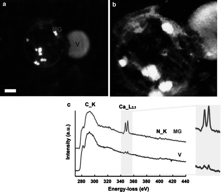
Analytical electron microscopic evidence of vesicle–mitochondrial interactions in mineralizing osteoblasts. a High-angle annular dark-field scanning TEM image of a dense granule-containing mitochondrion associating with a vesicle within an osteoblast in a mineralized nodule. The sample was prepared by high-pressure freezing and freeze substitution (HPF-FS). (Scale bar = 200 nm). b Voltex projection of a 3D tomographic reconstruction showing a mitochondrion conjoined with a vesicle. Dense granules are evident within the mitochondrion. Sample was prepared by HPF-FS. c Electron energy loss spectroscopy (EELS) of specified areas within the mitochondrion and vesicle in a. The mitochondrial granule and vesicle show characteristic calcium L2 and L3 edges at 346 eV. All spectra display carbon edges. d Orthoslices at 10-nm intervals through the tomographic reconstruction showing the mitochondrion–vesicle interface. The mitochondrial membrane is discontinuous where it conjoins the vesicle (arrows) [131]

