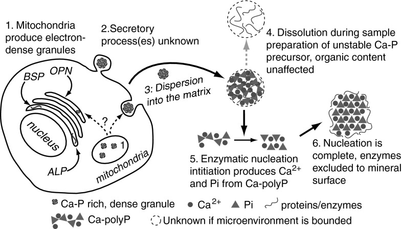
Fig. 4.
Proposed controlled apatite biomineralization schematic. (1) Mitochondria produce polyPs from phosphate sources that form complexes with calcium, producing discrete, electron-dense, Ca- and P-rich granules. (2) Granules may be processed through the trans-Golgi network (TGN), and then secreted via budding or exocytosis. If processed through the TGN, granules may associate with matrix and/or noncollagenous proteins as well as phosphatase enzymes. The secreted product is an amorphous Ca-/P-rich granule that contains matrix proteins. It is unknown if the granule is encapsulated. (3) The granule migrates to mineral nucleation sites within the collageneous matrix, where the noncollagenous proteins may play significant roles in granular interaction with the matrix. (4) During sample preparation, these unstable, amorphous granules may be artifactually dissolved so that only the remaining protein component is observed. These may be “crystal ghosts.” (5) If a phosphatase enzyme component of the unstable, amorphous precursor is activated within the matrix, the Ca–polyP component begins to transform into Ca2+ and Pi components. The local, high concentrations of Ca2+ and Pi nucleate apatite. As the apatite nucleus grows while the polyP depolymerizes, the protein component of the granule is excluded from the growing apatite crystal. This displaces the granule protein components to the surface. (6) The excluded proteins surround the apatite crystal surface, where they control crystal growth and shape, among other functions

