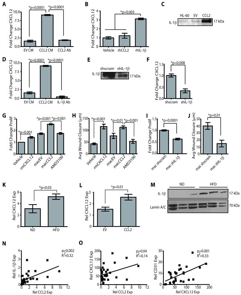
Figure 4. Macrophage derived CXCL12 induces angiogenesis.
(A) SVF/CCL2 conditioned media (CM) significantly increased CXCL12 expression in HL-60 macrophages compared with SVF/EV CM or SVF/CCL2 CM + blocking antibody for CCL2 (CCL2 Ab). (B) Recombinant human IL-1β (rhIL-1β) increased CXCL12 expression in HL-60 macrophages. qPCR was performed on RNA isolated from 3 experiments. (C) SVF/CCL2 CM contained increased IL-1β protein compared to SVF/EV CM. CM from HL-60 macrophages treated with LPS was used as a positive control. (D) SVF/CCL2 CM+IL-1β blocking Ab significantly reduced CXCL12 expression in HL-60 macrophages compared to SVF/CCL2 CM. (E) CM from SVF/CCL2 shIL-1β (shIL-1β) cells demonstrated decreased IL-1β protein compared to CM from SVF/CCL2 shscrambled control cells (shscram). (F) SVF/CCL2 shIL-1β CM significantly decreased CXCL12 expression in HL-60 macrophages compared with SVF/CCL2 shscramble CM. (G, H) Human microvascular endothelial cells (HMVEC) proliferated (G) and migrated (H) in response to recombinant mouse CXCL12 (rmCXCL12), as well as in response to CM from HL-60 macrophages pre-treated with CM from SVF/CCL2 cells (macCCL2) compared to those treated with SVF/EV CM (macEV). AMD3100+macCCL2 CM significantly decreased HMVEC proliferation and migration. Proliferation data are represented as a fold change of vehicle-treated cells, and 3 experiments were performed in triplicate. (I, J) HMVEC demonstrated decreased proliferation (I) and migration (J) in response to CM from HL-60 macrophages pre-treated with CM from SVF/CCL2 shIL-1β cells (mac shIL-1β) compared to CM from SVF/CCL2 shscrambled cells (mac shscram). Proliferation data are represented as a fold change of vehicle-treated cells, and 3 experiments were performed in triplicate. (K) Glands from C57Bl/6 HFD mice demonstrated elevated CXCL12 expression compared to glands from ND mice (n=6 mice/group). (L) SVF/CCL2 humanized glands from NOD/SCID mice demonstrated significantly increased CXCL12 expression compared to SVF/EV humanized glands (n=6 mice/group). (M) IL-1β protein was increased in C57Bl/6 HFD mammary glands compared to ND glands. (N, O) Reduction mammoplasty tissue demonstrated a significant correlation between CCL2 and IL-1β expression levels (N), as well as significant correlation between expression of CXCL12 and both CCL2 and CD31 (O) qPCR was performed on RNA isolated from 28 reduction mammoplasty samples.

