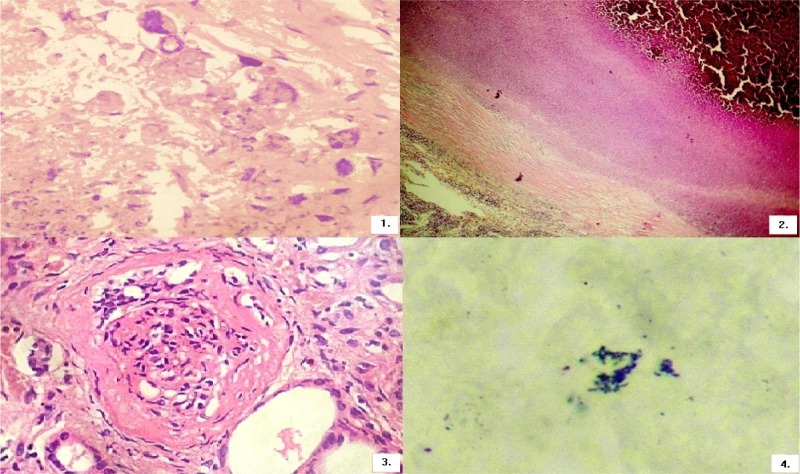
Figure 1.
PAS staining technique showing viable yeast cells suggestive of histoplasmosis.
Figure 2.
ZN staining is negative for tuberculosis.
Figure 3.
Mesangial sclerosis, cellular proliferation and thickened glomerular capsules in keeping with membrano-proliferative glomerulonephritis
Figure 4.
Grocots methenamine staining showing the typical morphological features of Histoplasma

