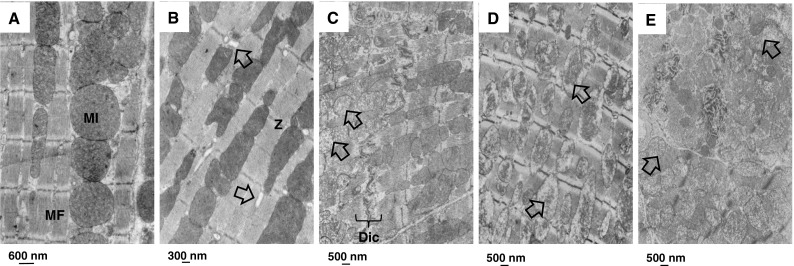
Fig. 8.

Mitochondrial changes in the myocardium after ErbB2 inhibition. Electron microscopic images of the myocardium of mice treated with lapatinib alone or in combination with doxorubicin at 40 weeks after treatment. a Age-matched control (n = 3); cardiomyocytes show organized sarcomeres characterized by parallel myofilaments anchored to Z bands and mitochondria were perfectly aligned and packed. b Doxorubicin treatment alone (n = 3); myofilament arranged regularly and mitochondria were aligned with focal vacuolization (arrow). c Lapatinib treatment alone (n = 3); focal damage per cardiomyocyte. Mitochondrial volume increased, mitochondrial cristae were fuzzy and had a cloudy swollen phenotype (arrow). d Direct lapatinib set up combined with doxorubicin (n = 3); e delayed lapatinib set up combined with doxorubicin (n = 3); disorganized mitochondria, mitochondrial cristae were fuzzy and had a cloudy swollen phenotype (arrow), increased volume of mitochondria. MI mitochondria; MF myofibril; Z z-bands; Dic Discus interculatis
