
Fig. 3.
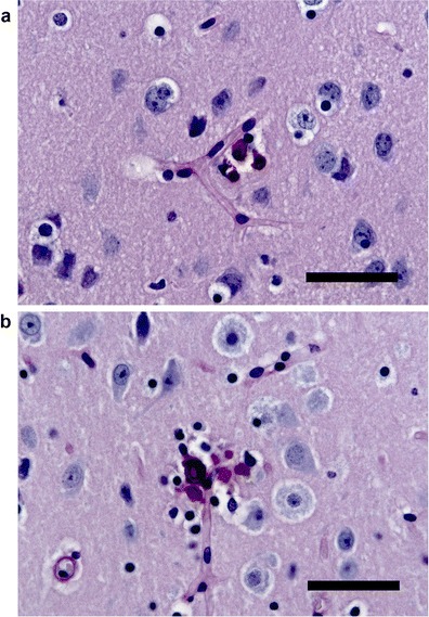
PAS staining of brain sections from homozygous Slc20a2 knockout mouse. Calcifications (purple stains) in close relation to blood vessels, delineated by the PAS-positive basal membrane, are found in the basal ganglia (a), thalamus (b), and cortex (not shown) (scale bars, 30 μm). No calcifications/lesions were observed in wt control (not shown)