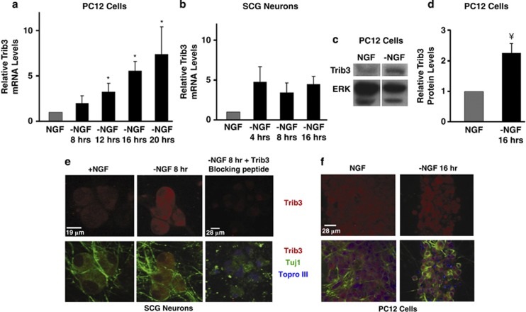
Figure 1.
Trib3 is induced in response to NGF deprivation. (a and b) Neuronal PC12 cells (a) and SCG neurons (b) were deprived of NGF for the indicated times. Total mRNA was isolated and subjected to reverse transcription followed by quantitative PCR using Trib3 primers. Rat GAPDH (glyceraldehyde 3-phosphate dehydrogenase) and α-tubulin mRNA levels were used to normalize input cDNA. The data are reported as relative increase in mRNA levels normalized to NGF control and represent mean±S.E. of three experiments for PC12 cells and±range of two experiments for SCG neurons. (c and d) Neuronal PC12 cells were deprived of NGF for 17 h. Whole-cell lysates were subjected to SDS-PAGE and western immunoblotted with anti-Trib3 and anti-ERK antisera. (c) Shows non-adjacent lanes from the same blot. (d) Shows quantification of Trib3 signal normalized to ERK signal. Values represent mean±S.E. for four independent experiments. (e and f) Immunocytochemistry of SCG neuron (e) and neuronal PC12 cell (f) cultures showing Trib3 protein upregulation after NGF deprivation. Note the absence of signal in the presence of a Trib3-blocking peptide. *P<0.05; ¥P<0.005 versus no NGF withdrawal (Student's t-test). Scale bar for SCG neurons=19 μm and for PC12 cells=28 μm

