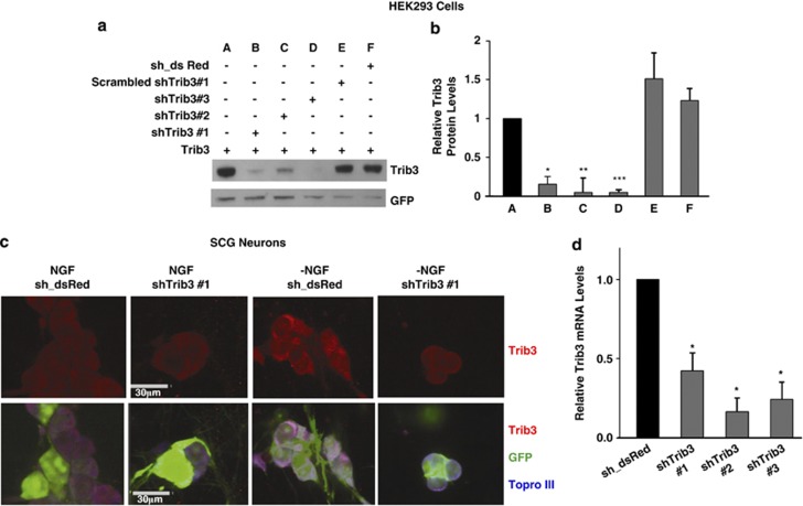
Figure 3.
shTrib3 constructs knock down both exogenous and endogenous Trib3. (a) HEK293 cells were co-infected with lentiviruses expressing Trib3 and shTrib3 or control shRNA constructs, and whole-cell lysates were prepared 3 days later and subjected to SDS-PAGE and immunoblotting for Trib3 or GFP (green fluorescent protein). (b) Quantification of relative Trib3 protein expression under conditions described in panel (a). Values are normalized to GFP expression to correct for infection efficiency and are expressed as means±S.E. of three experiments. Differences from Trib3 expressing samples without Trib3 shRNA: *P<0.005; **P<0.05; ***P<0.0005 (Student's t-test). (c) SCG neuronal cultures were infected with either control or shTrib3#1 expressing lentivirus for 3 days followed by 8 h of continued NGF treatment or NGF deprivation, all as indicated. Neurons were fixed and immunostained using antisera against Trib3. Bar=30 μm. (d) SCG cultures were infected with either control (sh_dsRed) or three different shTrib3 expressing lentiviruses for 3 days in the presence of NGF. Total mRNA was isolated and subjected to reverse transcription followed by quantitative PCR using Trib3 primers. Rat α-tubulin mRNA levels were used to normalize input cDNA. The data are reported as relative increase in mRNA levels normalized to NGF control and represent mean±S.E. of three experiments. *P<0.005 (Student's t-test) compared with control virus-infected neurons

