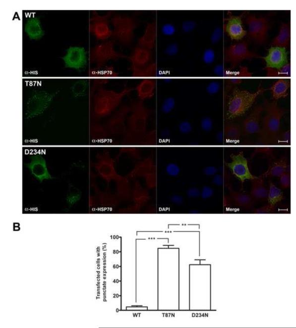
Figure 4.
Immunofluorescence microscopy from transiently transfected HEK-293 cells. A, Cells expressing recombinant HIS-CBS were detected by using mouse anti-polyhistidine antibody and goat anti-mouse Alexa 594 antibody (green). The cytoplasmic protein Hsp70 was immunodetected with rabbit anti-Hsp70 antibody and goat anti-rabbit Alexa 594 (red). The fourth unlabelled picture in each set is a merge of the other three images. Optical resolution 100X. Scale bar: 5 μm. B, Percentage of transfected cells with punctate expression of recombinant proteins. Results represent the average ± SEM of five independent experiments (***p< 0.001, **p< 0.01).

