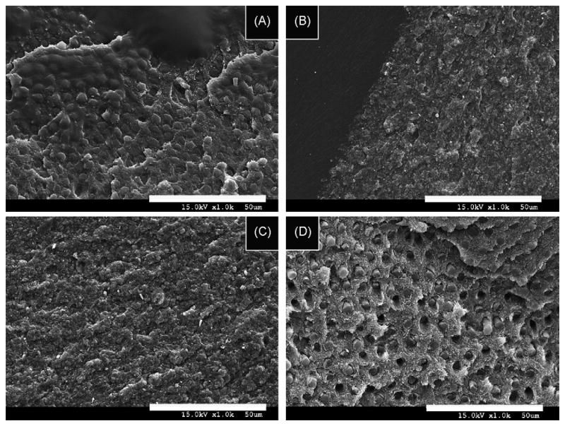
Fig. 2.

SEM micrographs of failure modes of hydrophilic resins applied to dentin conditioned with EDTA or PA. (A) SEM micrograph of the failure mode of resin-bonded dentin created with resin 3 applied to PA-etched dentin showing the bonded dentin failed in a mixed mode with the exposed dentin surface still covered by well-hybridized dentin, and no exposure of collagen fibrils or dentinal tubules. (B) SEM micrograph of the failure mode of resin-bonded dentin created with resin 3 applied on EDTA-treated dentin that failed in a mixed mode. The dentin surface remains covered with resin or hybridized dentin. No dentinal tubules are detected. (C) SEM micrograph of the failure mode of resin-bonded dentin created with resin 4 applied on EDTA-treated dentin. Note the absence of exposed dentinal tubules and the presence of resin on the dentinal surface. (D) SEM micrograph of the failure mode of resin-bonded dentin created with the most acidic and hydrophilic resin 5 applied on PA-etched dentin. Note the presence of fractured resin tags inside the dentinal tubules and collagen fibrils on the dentin surface left exposed by the cohesive failure of the hybrid layer. In the upper right portion of the image, the hybrid layer remains present. However, most of the image shows the top of the underlying sound dentin that was etched but not infiltrated.
