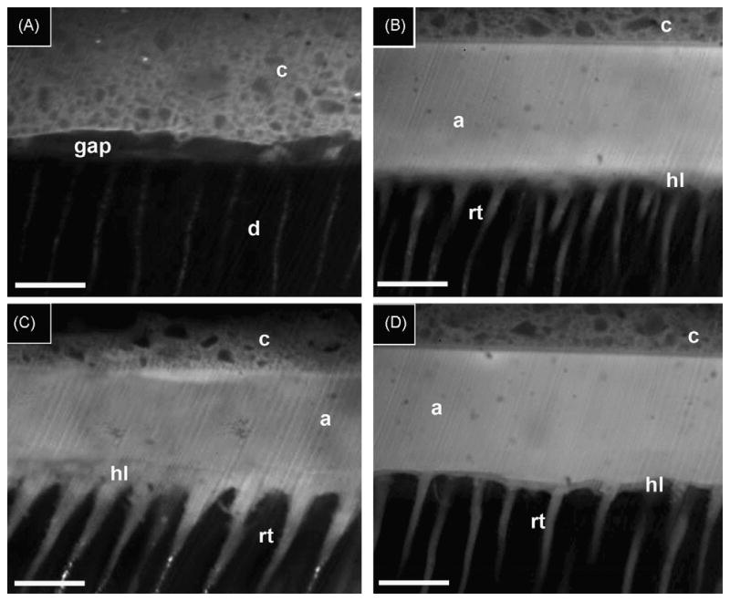
Fig. 4.

Confocal fluorescence of the resin–dentin interfaces. (A) Resin-bonded dentin interfaces created with hydrophobic resin 1 containing 0.1% rhodamine-B, applied on ethanol wet-dentin that was conditioned with EDTA for 60 s. Note that the absence of a hybrid layer and absence solid resin tags formation indicates that the primer and adhesive did not penetrate the dentin during the bonding procedures. Gaps were present between dentin (d) and composite (c). (B) Resin-bonded dentin interfaces created with resin 2 containing 0.1% rhodamine-B, applied on ethanol wet-dentin that was conditioned with EDTA for 60 s showed the presence of a very thin (ca. 1–2 t-m) hybrid layer (hl), the adhesive layer (a) and composite (c) layer. The scale bar in all the images is 10 t-m. (C) Resin-bonded dentin interfaces created with resin 2 containing 0.1% rhodamine-B, applied on acid-etched ethanol wet-dentin showed the presence of a thicker hybrid layer (hl) beneath the adhesive layer (a) and composite (c). The resin tags (rt) appeared to have larger diameters compared to those observed in the resin-bonded dentin interfaces created with resin 2 applied on EDTA/ethanol wet-dentin. (D) Resin-bonded dentin interfaces created with the primer and adhesive of SBMP containing 0.1% rhodamine-B, applied on ethanol wet-dentin that was conditioned with EDTA for 60 s. Note the presence of a thin (i.e. 1–2 t-m) hybrid layer (hl) and resin tags (rt) beneath the adhesive composite (c).
