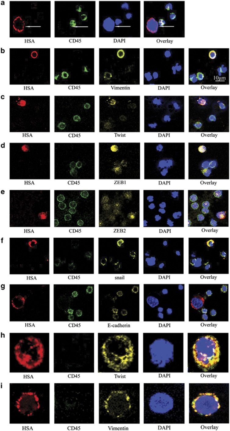
Figure 2.
Immunofluorescence staining identified the expression of EMT-related genes in CTCs obtained from HCC patients with different stages of disease. (a) CTCs and hematologic cells were stained with the HSA anti-mouse antibody/Alexa Fluor 647 rabbit anti-mouse IgG (red) and CD45 anti-rat antibody/Alexa Fluor 488 rabbit anti-rat IgG (green). The cell nuclei were stained with DAPI (blue). (b–g) Triple-immunofluorescence shows CTCs expressing vimentin, twist, ZEB1, ZEB2, snail and E-cadherin, respectively. These CTCs were stained with primary antibodies raised in rabbit against vimentin, twist, ZEB1, ZEB2, snail and E-cadherin, and the corresponding secondary antibody was Alexa Fluor 555 donkey anti-rabbit IgG (yellow). The remaining staining procedures were as same as those described for double-immunofluorescence. Bar=10 μm. Twist and vimentin coexpression in CTCs obtained from the same patient. Representative photomicrographs of CTCs obtained from the same patient in different visual fields. (h) A CTC expressing HSA and twist but not CD45. (i) A CTC expressing HSA and vimentin but not CD45. Original magnification × 800

