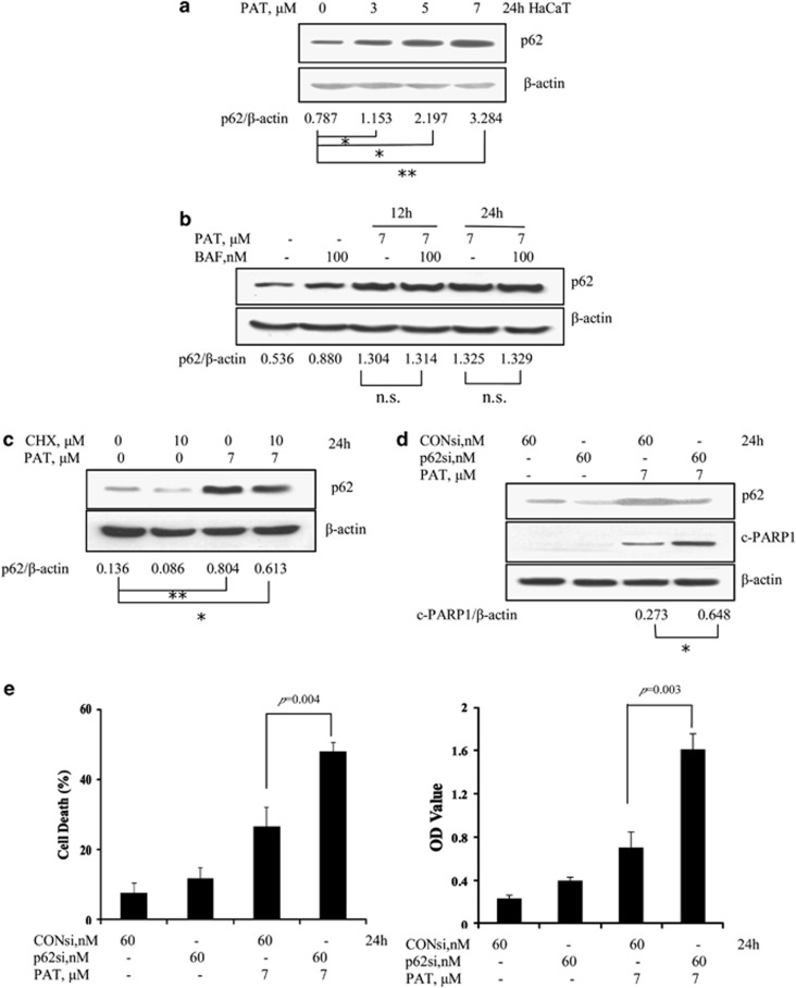
Figure 2.
Inhibition of autophagosome degradation-mediated induction of p62 protects PAT-induced apoptosis in HaCaT cells. (a) p62 accumulation induced by PAT in HaCaT cells. The cells were treated with various concentrations of PAT for 24 h and then p62 protein levels were analyzed by western blotting. (b) Effects of autophagosome degradation inhibition by BAF on PAT-induced accumulation of p62. The cells were treated with 7 μM PAT for 12 and 24 h in the presence or absence of 100 nM BAF (added 2 h before cell harvest) and then p62 was analyzed by western blotting. (c) Effects of protein synthesis inhibition by CHX on PAT-induced accumulation of p62. The cells were treated with 7 μM PAT for 24 h in the presence or absence of CHX and then p62 was analyzed by western blotting. Effects of p62 inhibition by RNAi on PAT-induced PARP1 cleavage (d) and apoptosis (e). The cells were transfected with 60 nmol/l of p62 siRNA using siPORT NeoFX transfection agent. At 24 h post transfection, the cells were treated with 7 μM PAT for 24 h. PARP1 cleavages were analyzed by western blotting and cell death was assessed by Annexin V staining (left) and a Cell Death Detection ELISA kit (right). n=3, *P<0.05, **P<0.01, n.s., no significant difference

